Pictures and Videos of Parasites
Pictures |
||||||
Only one bot fly species attacks humans, the Dermatobia hominis. From Wikipedia: Botflies deposit eggs in a host body, or sometimes use an intermediate vector: common houseflies for example. Eggs are deposited in animal skin directly, or the larvae drop from the egg: the body heat of the animal induces hatching upon contact. Some forms of botfly also reside in the digestive tract when consumed by a licking action. Myiasis can be caused by larvae burrowing into the skin (or tissue lining) of the host animal. Mature larvae drop from the host and complete the pupal stage in soil. |
||||||
Ascaris eggs are found in human feces. After feces contaminates the soil, the eggs become infectious after a few weeks. Infection occurs when a person accidentally ingests (swallows) infectious Ascaris eggs. Once in the stomach, immature worms hatch from the eggs. The larvae are carried through the lungs and then to the throat where they are swallowed. Once swallowed, they reach the intestines and develop into adult worms. Adult female worms lay eggs that are then passed in feces; this cycle will take between 2-3 months. Pigs can be infected with another species of Ascaris. Occasionally, a pig Ascaris infection can be spread to humans; this occurs when infective eggs, found in the soil and manure, are ingested. Infection is more likely if pig feces is used as fertilizer in the garden; crops then become contaminated with Ascaris eggs. You or your children can become infected after touching your mouth with your hands that have become contaminated with eggs from soil or other contaminated surfaces or by ingesting contaminated food or water. |
||||||
However, the actual story is that it was a man in his 70s who was suffering from an unusual form of cancer which had eaten away at the upper portion of his skull and scalp but had not caused him any pain so he had not sought medical help. He was involved in a minor auto accident and the paramedics found him in this condition. There was no explanation as to whether a minor accident had caused the damage and dislodged fragile tissue or if the scalp had already been eaten away. There were indeed many insects and maggots infesting his brain. |
||||||
Onchocerciasis is the world's second leading infectious cause of blindness. Rarely life-threatening, the disease causes chronic suffering and severe disability. In Africa, it constitutes a serious obstacle to socioeconomic development. It is often called river blindness because of its most extreme manifestation and because the blackflies that transmit the disease abound in riverside areas, where they breed in fast-flowing waters. Fertile riverine areas are frequently abandoned for fear of the disease. The disease is caused by Onchocerca volvulus. It is mostly found in Africa but also a few countries in Latin America. A parasitic worm, Onchocerca volvulus, of the Adult worms remain in subcutaneous nodules, limiting access to the host's immune system. Microfilariae, in contrast, are able to induce intense inflammatory responses, especially upon their death. Dying microfilariae have been recently discovered to release Wolbachia-derived antigens, triggering innate immune responses and producing the inflammation and its associated morbidity. Wolbachia species have been found to be endosymbionts of O. Volvulus adults and microfilariae and are thought to be the driving force behind most of O. Volvulus morbidity. Severity of illness is directly proportional to the number of microfilariae and the power of the resultant inflammatory response. |
||||||
Guinea Worms. First swallowed by a "Cyclops" or water flea, the Guinea worm transforms into a third stage larvae. The Cyclops, ingested by a human being, are consumed by the stomach's juices and the larvae of the Guinea Worm are released. They remain in the stomach for up to three months. After mating, the male dies and the female bores through the body making her way to the extremities, usually the lower leg or foot, but she can go to any part of the body. Once settled, just under the skin, she begins to grow, by eating the flesh of her carrier, into a three to As the worm matures, a painful blister appears on the skin of the carrier. When the person puts the affected part of the body in water, the blister breaks and hundreds of thousands of tiny first stage larvae are released into the water. The adult female worm then comes slowly out of the body of its carrier through the sore made by the broken blister. It usually takes several weeks for the worm to completely exit the body. People, having no choice, drink the infested water, ingest the worm and the entire cycle begins once again. No matter how often people are infected they do not become immune to the Guinea Worm. Both young and old alike are subject to this dreaded parasite. |
||||||
Demodex folliculorum, or the demodicid also known as the eyelash mite, is a tiny mite, less than 0.4 mm long, that lives in pores and hair follicles, usually on the nose, forehead, cheek, and chin, and often in the roots of eyelashes. (A follicle is the pore from which a hair grows). It is not really a parasite since it lives on dead skin cells and secretions, but overgrowth of them can cause irritation.
The mites live head-down in a follicle, feeding on secretions and dead skin debris. At the left, you can see seven demodicids buried in the follicle of a hair, and you can also see the hair's shaft. If too many mites have buried into the same follicle, it may cause the eyelash to fall out easily. An individual female may lay up to 25 eggs in a single follicle, and as the mites grow, they become tightly packed. When mature, the mites leave the follicle, mate, and find a new follicle in which to lay their eggs. The whole cycle takes between 14 to 18 days. Sometimes demodex is called the 'face mite', since it is often associated with blackheads, acne and other skin The mites have tiny claws, and needlelike mouthparts for eating skin cells. Their bodies are layered with scales, which help them anchor themselves in the follicle. The mite's digestive system results in so little waste that, unlike the dust mite, this mite doesn't even have an excretory opening. So although there may be mites in your eyelashes, there isn't any mite poop! Thank goodness! However ... did you know that you go to sleep at night on a pillow that is home to many thousands of dust mites ...which help keep our homes clean by consuming the tens of millions of skin cells we shed each day? Just pretend they're not there! |
||||||
There are numerous species of tapeworm, which may be as long as 50 ft. (15 m). Their bodies are made up of segments called proglottids, which contain male and female sexual organs and which house their eggs. The Taenia genus of tapeworm includes T. solium, the pork tapeworm, which has been known to crawl out of the anus of an infected human. Another worm often found in under-cooked pork is Trichinella spiralis, which brings about the condition known as trichinosis. The latter disease is rarely fatal, but it can cause extreme discomfort, characterized by sore, tender muscles. Thanks to enhanced efforts at meat inspection, the incidence of trichinosis in U.S. pigs has dropped to less than 1%. Nonetheless, it is still quite possible to ingest the parasite from eating undercooked game, particularly bear. |
||||||
|
||||||
|
||||||
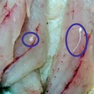
From Florida Fish and Wildlife Research Institute - Imagine a day fishing out on the water and everything seemed great. A legal limit of good-sized fish has been caught, plenty for a tasty dinner upon returning home. But while filleting one of the fish, there are spots found in the muscle, or even visible worms. What is one to do, cut out those areas and continue to cook and enjoy the fish for dinner, or throw it away? Although these parasites may not look very appetizing, worms are commonly found in marine fish and are rarely passed from fish to humans. It is safe to cut away the affected area and continue to cook the fish as usual. Even if parasites are present cooking kills them and they are not a risk to public health.
Parasites are organisms that live on or within another organism, called the “host.” Most fish species are susceptible to worm infestations but a few species, particularly those in sharks and fishes in the grouper, amberjack, and drum families, appear to be more susceptible to them. Larger, older fish can acquire many parasites over the course of their lives. Though they appear to be very intrusive, parasites rarely cause health complications for the fish. Although most commonly seen by anglers in the muscle, parasitic worms can live in every organ. For example the ovary of this grouper is inhabited by large, red nematodes or “roundworms.” In general, parasite density and diversity can be good indicators of the overall health of a marine environment. Fish possessing a high diversity of species of parasites in relatively low abundances are commonly found in healthy marine systems. Also see the article Fish and Parasites |
||||||
Lymphatic filariasis is a parasitic disease caused by microscopic, thread-like worms. The adult worms only live in the human lymph system. The lymph system maintains the body's fluid balance and fights infections. Lymphatic filariasis is spread from person to person by mosquitoes. People with the disease can suffer from lymphedema and elephantiasis and in men, swelling of the scrotum, called hydrocele. Lymphatic filariasis is a leading cause of permanent disability worldwide. Communities frequently shun and reject women and men disfigured by the disease. Affected people frequently are unable to work because of their disability, and this harms their families and their communities. |
||||||
From Dermatology Info.net : Cutaneous leishmaniasis has many local synonym such as Tropical sore, Children are more susceptible, where solid immunity is acquired after the first infestation. This is why some natives sometimes inoculate their children with the protozoa on the shoulder or thighs to have the disease there in order to protect the face from scarring if they are infested in the future with leishmania.
Picture at left: Surrounded by the forest-like hairs of her victim, a sand fly delivers what could be a parasite-carrying bite that causes leishmaniasis. Fall is peak sand fly season in Iraq, where service members who do not follow protective measures could be at risk for the disease. In 2002 and 2003, 73 service members were diagnosed with the cutaneous (skin) version of the disease. All but two spent time in Iraq. |
||||||
Ascaris is a worm that lives in the small intestine. Infection with Ascaris is called ascariasis (ass-kuh-rye-uh-sis). Adult female worms can grow over 12 inches in length, adult males are smaller. Ascariasis is the most common human worm infection. Infection occurs worldwide and is most common in tropical and subtropical areas where sanitation and hygiene are poor. Children are infected more often than adults. In the United States, infection is rare, but most common in rural areas of the southeast. Most people have no symptoms that are noticeable, but infection may cause slower growth and slower weight gain. If you are heavily infected, you may have abdominal pain. Sometimes, while the immature worms migrate through the lungs, you may cough and have difficulty breathing. If you have a very heavy worm infection, your intestines may become blocked.
Pigs can be infected with another species of Ascaris. Occasionally, a pig Ascaris infection can be spread to humans; this occurs when infective eggs, found in the soil and manure, are ingested. Infection is more likely if pig feces is used as fertilizer in the garden; crops then become contaminated with Ascaris eggs. You or your children can become infected after touching your mouth with your hands that have become contaminated with eggs from soil or other contaminated surfaces or by ingesting contaminated food or water. |
||||||
|
||||||
Videos |
||||||
Comments on video of  ascaris removal from duodenum: This 34 year old female resident of rural China presented with nausea and vomiting, vague abdominal pain and fever. An upper endoscopy was performed in Beijing. As the endoscope is advanced to the second portion of the duodenum (the duodenum is the start of the small intestine which is attached to the stomach), a live Ascaris was encountered. Ascaris infections occur worldwide, with current estimates suggesting that 1.4 billion people may be infected with this parasite. The roundworm is retrieved using a biliary basket. Once the worm is secured in the basket, both worm and endoscope were removed from the patient. Notice here the length of the organism, about 20cm in total, and the continued movement even after it is removed. ascaris removal from duodenum: This 34 year old female resident of rural China presented with nausea and vomiting, vague abdominal pain and fever. An upper endoscopy was performed in Beijing. As the endoscope is advanced to the second portion of the duodenum (the duodenum is the start of the small intestine which is attached to the stomach), a live Ascaris was encountered. Ascaris infections occur worldwide, with current estimates suggesting that 1.4 billion people may be infected with this parasite. The roundworm is retrieved using a biliary basket. Once the worm is secured in the basket, both worm and endoscope were removed from the patient. Notice here the length of the organism, about 20cm in total, and the continued movement even after it is removed. |
||||||
Comments on video of removal of anisakis simplex seen during endoscopy: This 61 year old restaurant owner was referred for upper endoscopy to evaluate typical longstanding dyspepsia and reflux type symptoms. His upper endoscopy appeared quite normal except for what initially appeared to be a wisp of mucin floating in the gastric fundus. On closer inspection, this worm like structure was seen and identified as the marine parasite anisakis simplex which is the cause of anisakiasis.
If the host dies, the larva migrate to the muscle tissues where through predation, they will pass onto another host. If instead they are consumed by marine mammals, they will mature in the host's intestine to shed eggs as adults. Humans become incidental hosts when they consume raw fish often in the form of sushi. The larvae hook into the gastric or intestinal wall and can cause a significant localized reaction and in some case peripheral eosinophilia. Most often, the clinical course following ingestion is asymptomatic: the larva briefly anchoring to the gastric or intestinal wall before dying and passing uneventfully. Larva have been reported to penetrate the mucosa and cause abdominal pain and vomiting. Small bowel lesions mimicking Crohn's disease have been noted. When seen endoscopically, removal as demonstrated in this case is curative. Generally, medical therapy is not necessary, but limited experience with albendazole has been reported. |
||||||
Comments on video of ascaris seen during colonoscopy: On screening colonoscopy, this abnormality was encountered in the cecum. This round worm is Ascaris Lumbricoides, one of the most common human parasites in the world. When ingested, the durable Ascaris eggs hatch in the small intestine releasing larva that migrate through the intestinal wall, and travel both hematogenously and lymphatically to the heart and lungs. Over the next several days, the larva mature in the alveoli, then migrate up the trachea to be swallowed back into the gastrointestinal The majority of Ascaris infections are as in this example asymptomatic. Symptoms are a consequence of either the immunologic hypersensitivity of the host to the worm as in the pulmonary stage referred as Loffler's syndrome or to mechanical obstruction of lumen by the worm. Heavy worm burden can result in intestinal obstruction and migrating worms can cause pancreatitis and/or cholangitis when involving the pancreatobiliary tree. Multiple medical therapies are approved for its treatment including mebendazole. Epidemiologically, infections are most common in areas of lower socio-economic conditions. This man manages a pig farm in China that is used to test pharmaceutical agents. From an endoscopic standpoint it is noteworthy that the worms do not like light and will move away fro the attention it is receiving. In this example, the endoscopist was too slow to snare his prey which succeeded in escaping temporarily into the cooler and darker confines of the small bowel out of reach of the endoscope but not from the soon to be consumed anti-helminthic therapy. |
||||||
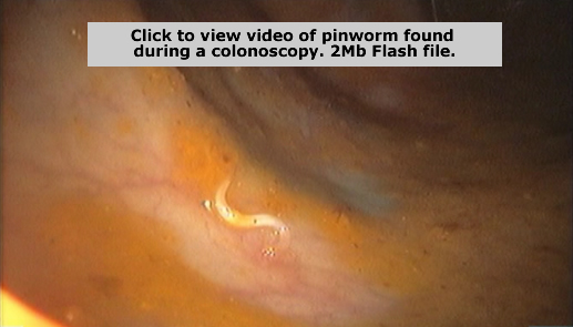
Comments on video of pinworm seen during colonoscopy: This 52 year old woman was referred for |
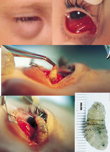 Doctors in Honduras removed a 2 centimeter long botfly larva from a child's eye socket.
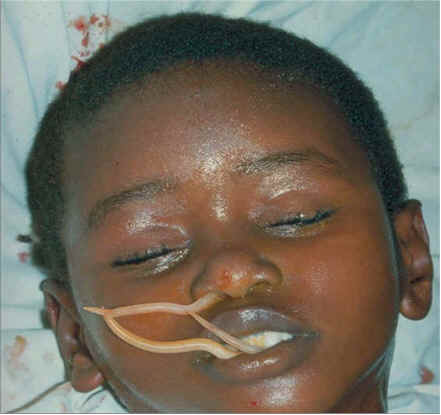 Severe ascaris infection with worms exiting through mouth and nose. Anesthesia appears to agitate the worms and when there is infection of adults in the lungs they can exit the mouth and nose. The normal life cycle of ascaris is to migrate to the lungs for the larval stage and grow to an adult in the intestines, but sometimes the adults reside in the lungs.
 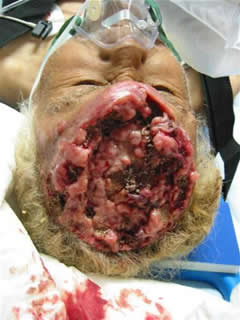 These two pictures have floated around the internet with the explanation that it was of an avid sushi eater who got some infestation which infected his brain. When he went to the doctor complaining of headaches, he was sent for surgery and this is
 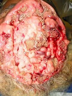 what was found. Some tellings of the story report that it is a form of tapeworm with many eggs, and others that maggots somehow got under his scalp and ate his brain.
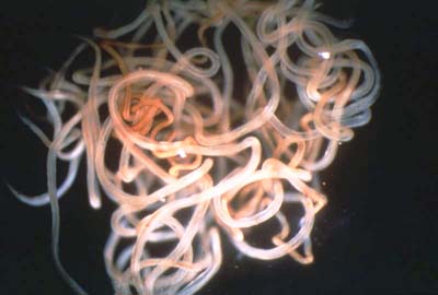
 family filariidae, lives in the human body for up to 14 years. Rarely life-threatening, the disease causes chronic suffering and severe disability. The male is 2-3 cm long and the female is up to 60 cm long. The vector for this parasite is the blackfly.
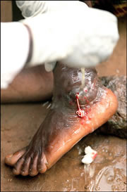

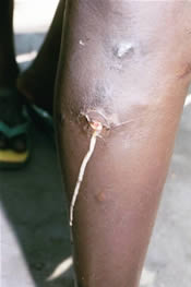
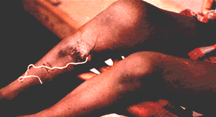 five foot long worm. This worm is about as big around as a piece of spaghetti. While growing, she causes severe pain and cripples the carrier, so that they are not able to work. These are simple subsistence farmers. Pain hinders their farming, causing them to survive on less and keeps their family from eating!
  Demodicids have a wormlike appearance, with legs that are mere stumps. People with oily skin, or those who use cosmetics heavily and don't wash thoroughly, have the heaviest infestations ... but most adults carry a few demodicids. Inflammation and infection often result when large numbers of these mites congregate in a single follicle.
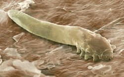 disorders (although it is not the cause of these). Demodex are harmless and don't transmit diseases, but large numbers of demodex mites may cause itching and skin disorders, referred to as Demodicosis.
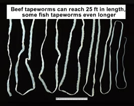 Proglottids break off, usually into the feces of the host, thus enabling the spread of the tapeworm. One variety, Dipylidium caninum (cucumber tapeworm), uses dogs or cats as a definitive host, entering the body when the animal ingests a flea or louse, which serves as an in
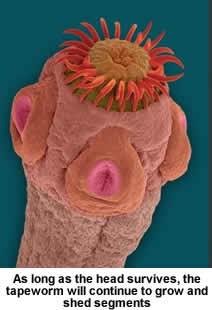 termediate host. Several varieties of worm can enter the body through improperly cooked pork. This is a testament to the wisdom of the injunctions against eating pork in the Old Testament and Koran, which were written for peoples in a world without refrigeration or sophisticated medical knowledge.
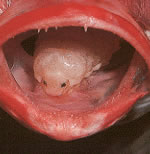 From
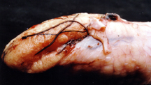
From 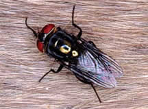 The screwworm is a type of fly but the larva growing from the eggs is what causes problems in humans and animals. It is likely that many of the "maggots" in the brain picture above are screwworm larvae. From
 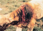 animal (usually some sort of livestock, but an injured soldier or a human baby isn’t out of the question) in search of a place to lay her eggs. She prefers wounds, but may also settle on using the eyes, nostrils, or anus of her victim to construct a nursery. Next, the 200-or-so eggs hatch, and the larvae start burrowing into their host’s flesh. Once they’re situated in their cozy little meat tunnels, the infant flies continue to feed and grow. The bigger they get, the more they have to eat. Eventually, this creates a whole lot of festering and oozing on the host, which attracts more flies, which lay more eggs, which do more feeding and burrowing. It’s a brutal onslaught, and a swift one. Screwworm larvae are reportedly capable of consuming an entire sheep or dog from the inside out in five to seven days.
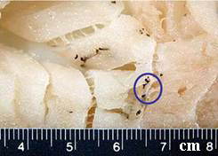
 Elephantiasis, or Lymphatic Filariasis, is a rare disorder of the lymphatic system caused by parasitic worms such as Wuchereria bancrofti, Brugia malayi, and B. timori, all of which are transmitted by mosquitos. Inflammation of the lymphatic vessels causes extreme enlargement of the affected area, most commonly a limb or parts of the head and torso. It occurs most commonly in tropical regions and particularly in parts of Africa.
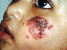 Oriental sore, Aleppo sore or Baghdad sore. The disease is caused by Leishmania tropica protozoa, which is endemic in Asia minor, Southwest Asia, the Mediterranean and gulf regions.The Phlebotomus sand fly is the vector, transmitting the disease from the reservoirs to human being. Direct infection from infected sores to a traumatized skin may rarely cause the disease .
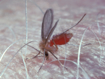 The disease has a very chronic course. The incubation period may take from weeks to two months from the beginning of the sand fly bite. Leishmaniasis usually affects children more than other age groups where the face, extremities and the neck are the most common sites involved.
 Massive ascaris infection in child shown at right. A large bolus of worms was expelled following antihelminthic treatment. This is one of the best-known photographs showing severe roundworm infection.
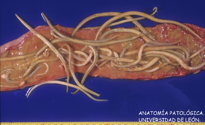 Ascaris eggs are found in human feces. After feces contaminates the soil, the eggs become infectious after a few weeks. Infection occurs when a person accidentally ingests (swallows) infectious Ascaris eggs. Once in the stomach, immature worms hatch from the eggs. The larvae are carried through the lungs and then to the throat where they are swallowed. Once swallowed, they reach the intestines and develop into adult worms. Adult female worms lay eggs that are then passed in feces; this cycle will take between 2-3 months.
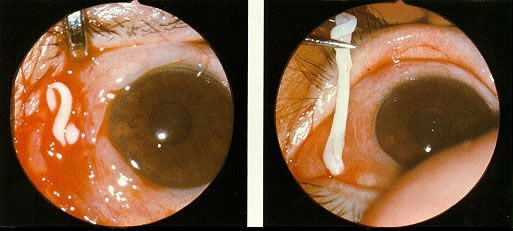 Removal of Sparganosis mansoni, a tapeworm larva, from eye. Adult tapeworms can reach lengths of 100cm or 40 inches. After penetrating the mucosa lining of the small intestine, they migrate systemically throughout the body, invading a variety of tissues and organs, and can live for years.

Links Electroherbalism's Antiparasitic General Regimen is an article in the Regimens section. Intestinal Roundworms, the Silent Killers is a humorous short video done by Michael Deash, Mike Baker, and Josh Clarke. Well, actually it is pretty dumb but then there's not much other parasitic roundworm humor out there. Ro-Revus Talks About Worms is a "serious" but unintentionally funny video of a gravelly-voiced frog puppet named Ro-Revus explaining intestinal worms. This video from 1971 was produced by The Malnutrition and Parasite Project of the Univerisity of South Carolina and funded by the Office of Economic Opportunity. A colonscopy video of a large pinworm infection. Fish and Parasites is an article in Electroherbalism's Parasite section. The CDC Parasitology website has some good jpg pictures in the "Identification and Diagnosis of Parasites of Public Concern" section. They include for many parasites diagnostic findings with microscopy *and* for some of the larger bugs, macroscopy, which is what they look like in their "natural" state instead of mounted and dyed like most parasite pics are. They also have a great section on "Morphologic comparisons of intestinal parasites" for help in diagnosing parasites from stool samples. Parasite Image Library . Provides multiple views plus pictures of life cycles for many parasites. Geneva Diagnostics, formerly known as The Great Smokies Diagnostic Laboratory, now known as Geneva Diagnostics, was the first large conventional lab diagnostic testing facility in the US to recognize the significance of pathogens like yeast and parasites and is still the most renowned. They also do metabolic diagnostics. Cells Alive! is a site dealing mostly with microscopic pathogens and explains in a simple way some aspects of cell biology plus includes many videos of the processes. It has a short pictorial article on how a virus (bacteriaphage) can infect bacteria (e. coli) and immunology topics like allergies, how mites cause itching, antibody production, and even the anatomy of a splinter Skin Manifestations of Medical Helminthes explains parasites which are of dermatological interests since they are associated with visible symptoms of the skin. This is a chapter from Principles of Pediatric Dermatology, an illustrated online book which contains much valuable information on many types of dermatitis, skin infections, and other dermatological topics. |